Fluorescein Angiography
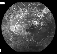
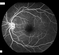 Fluorescein angiography is a diagnostic test that involves taking photographs of the blood vessels in the eye with the help of a contrast dye. Fluorescein is a yellow dye that allows the eye to glow using a certain wavelength of visible light. The images produced by this test helps doctors evaluate the retina and diagnose or monitor problems such as diabetic retinopathy, macular degeneration, abnormal vessel growth, macular edema, retinal artery or vein occlusions, certain types of retinal detachment, and tumors.
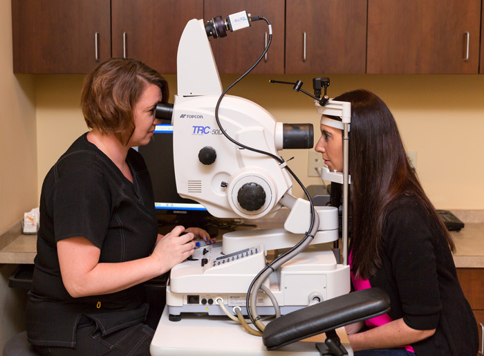
During these exams, the patient’s pupils are dilated with eye drops. A few photographs are then taken with a special ophthalmic camera. Next, the contrast dye is injected, usually into a vein in the patient’s arm. The dye travels up to the eye within a few seconds and “lights up” the blood vessels for the camera. Once the dye is injected, multiple photographs are taken.
These tests are considered safe for most patients, although it is possible to have a reaction to the dye and develop itching, nausea and a rash. These symptoms can usually be managed through oral medication if severe. In addition, patients who receive fluorescein injections can expect that their urine will appear bright yellow-orange for the first several hours after the test.
 Fluorescein dye is considered safe for most patients since it is a vegetable-based dye. It does not contain iodine and can generally be used even in patients who have had reactions to other intravenous contrast dyes used by radiologists and other specialists. Fluorescein angiograms may be required intermittently to assess a patient’s response to treatment or need for further therapy.
Recent Comments