Optical Coherence Tomography
Optical coherence tomography (OCT) is an advanced technology used to produce cross-sectional images of the retina, the light-sensitive lining on the back of the eye where light rays focus to produce vision. These images can help with the detection and treatment of serious eye conditions such as macular holes, macular puckers, and retinal swelling from diabetic retinopathy or macular degeneration.
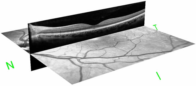
OCT uses technology that is similar to CAT scans of internal organs, using a scattering of light to rapidly scan the eye to create an accurate cross-section. Unlike other imaging techniques, OCT uses light to produce high resolution images, rather than radiation, sound, or radiofrequency waves. As a result, there is no risk to the patient or harmful side effects.
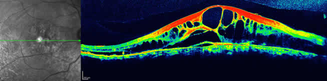
Your doctor can evaluate and measure each layer of the retina through this image and compare it with normal, healthy images of the retina. This technology allows your doctor to examine the retina at a microscopic level and has revolutionized the diagnosis and treatment of many retinal conditions. OCT scans may be performed multiple times during the course of treatment to assess both response to therapy and need for further intervention.
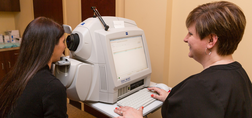
The OCT exam is non-invasive and usually takes about 10 minutes to perform in your doctor’s office.
Recent Comments